A comparison of synchrotron X-ray phase contrast tomography and holotomography for non-invasive investigations of the internal anatomy of mites
Keywords:
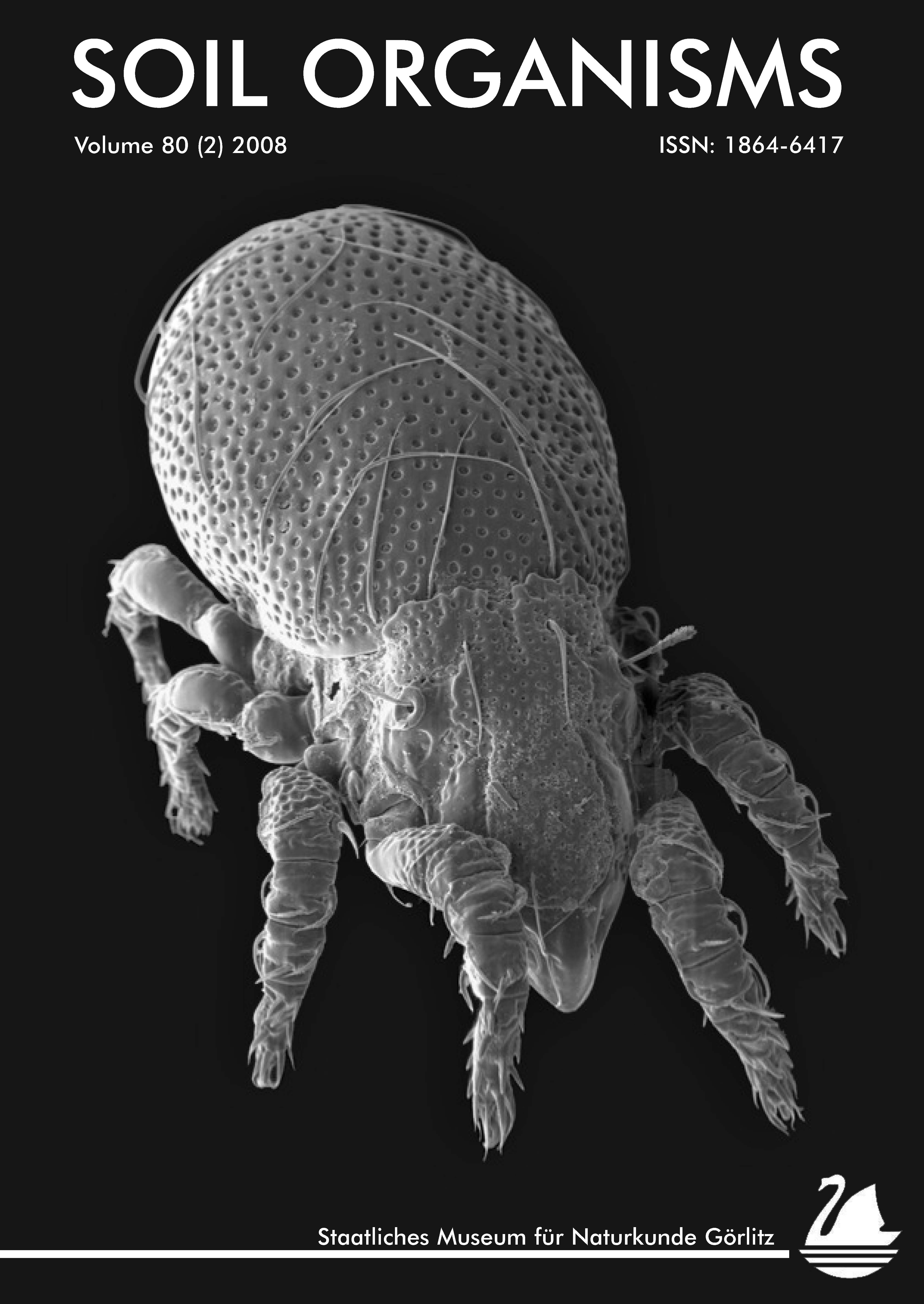
Acari, Oribatida, μCT, X-ray imagingAbstract
Synchrotron X-ray tomography is a powerful tool for non-invasive studies of the internal anatomy of microarthropods. The invention of phase contrast imaging (PCT) enables the visualisation of biological tissues with a small range of attenuation coefficients. Quantitative phase tomography (holotomography; HT) is an advancement of PCT and improves the imaging quality of materials with even smaller differences in attenuation coefficients. In this study, HT was used for the first time to investigate the internal anatomy of microarthropods. Both techniques, HT and PCT, are compared with respect to their ability to differentiate between soft tissues with low attenuation coefficients of the oribatid mite Archegozetes longisetosus (Acari, Oribatida). HT has a higher signal-to-noise ratio and a broader grey value distribution and resolves slight variations in soft tissues much better than PCT
Downloads
Published
How to Cite
Issue
Section
License
All articles from Senckenberg’s SOIL ORGANISMS Open Access scientific journal that are made available on the Senckenberg website (www.senckenberg.de) and also www.soil-organisms.org may be read, copied, distributed, and (in limited quantity) printed for non-commercial, private, scientific purposes.
In accordance with the German Science Foundation’s „Rules for the Safeguarding of Good Scientific Practice“, references to cited articles are to be complete and correct and furnished with a link to the website of the Senckenberg journal in question.
The Senckenberg Society for Nature Research (Senckenberg Gesellschaft für Naturforschung, SGN) is a member of the Leibniz Association (Leibniz-Gemeinschaft) and is therefore committed to the idea of Open Access as explained in the Berlin Declaration (Berlin Declaration on Open Access to Scientific Knowledge, Berliner Erklärung über den offenen Zugang zu wissenschaftlichem Wissen).
Open Access is understood to mean the charge-exempt public access to scientific results via the internet. The users should be able to read, copy, print, search within, and reference the full text without limitation and to use it in any conceivable lawful manner without financial, legal or technical hindrance.
This applies also to the SGN, which publishes various scientific series. Some scientific journals are made available to the public via Open Acess in addition to printed copies.
