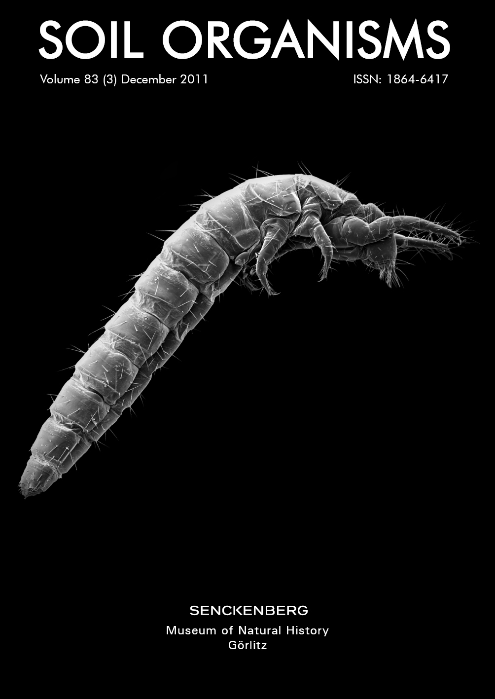
Confocal imaging of the exo- and endoskeleton of Protura after nondestructive DNA extraction
Keywords:
Cuticle, CLSM, autofluorescence, Congo redAbstract
In certain minute arthropods, such as Protura, species determination cannot be performed unambiguously without clearing and slide mounting of specimens. This causes an awkward dilemma for scientists conducting molecular research, since conventional DNA extraction entails destruction of the whole specimen. Thus, single individuals can be used either to obtain molecular data or for determination purposes. Such molecular datasets are thus dependent on determination of co-habitant specimens, and entries in GenBank are highly prone to misidentification.
To overcome this problem, we applied a non-destructive DNA extraction method and subsequently used confocal autofluorescence imaging to analyse and document cuticular characters of the same specimens. Alternatively the preparations can be examined by conventional microscopy. Our results show that the used non-destructive extraction method results in completely clear cuticular remains and does not significantly affect autofluorescence or shape. The acquired confocal image stacks and resulting volume renderings are useful to visualise, reconstruct and quantify structures for taxonomic purposes but also for morphological investigation of special cuticular structures such as the head endoskeleton of hexapods.
Downloads
References
Christian, E. & A. Szeptycki (2004): Distribution of Protura along an urban gradient in Vienna. – Pedobiologia 48: 445–452.
Dell’Ampio, E., N. U. Szucsich, A. Carapelli, F. Frati, G. Steiner, A. Steinacher & G. Pass (2009): Testing for misleading effects in the phylogenetic reconstruction of ancient lineages of hexapods: influence of character dependence and character choice in analyses of 28S rRNA sequences. – Zoologica Scripta 38(2): 155–170.
Difato, F., F. Mazzone, S. Scaglione, M. Fato, F. Beltrame, L. Kubínová, J. Janácek, P. Ramoino, G. Vicidomini & A. Diaspro (2004): Improvement in volume estimation from confocal sections after image deconvolution. – Microscopy Research and Technique 64: 151–155.
François , J., R. Dallai & W. Y. Yin (1992): Cephalic anatomy of Sinentomon erythranum Yin (Protura: Sinentomidae). – International Journal of Insect Morphology and Embryology 21(3): 199–213.
Gilbert, M. T. P., W. Moore, L. Melchior & M. Worobey (2007): DNA extraction from dry museum beetles without conferring external morphological damage. – PLoS ONE 2(3): e272.
Hall, T. A. (1999): Bioedit: a user-friendly biological sequence alignment editor and analysis program for windows 95/98/nt. – Nucleic Acids Symposium Series 41: 95–98.
Klaus, A. V., V. L. Kulasekera & V. Schawaroch (2003): Three-dimensional visualization of insect morphology using confocal laser scanning microscopy. – Journal of Microscopy 212: 107–121.
Klaus, A. V. & V. Schawaroch (2006): Novel methodology utilizing confocal laser scanning microscopy for systematic analysis in arthropods (Insecta). – Integrative and Comparative Biology 46(2): 207–214.
Lee, S., R. L. Brown & W. Monroe (2009): Use of confocal laser scanning microscopy in systematics of insects with a comparison of fluorescence from different stains. – Systematic Entomology 34: 10–14.
Michels, J. (2007): Confocal laser scanning microscopy: using cuticular autofluorescence for high resolution morphological imaging in small crustaceans. – Journal of Microscopy 227(1): 1–7.
Michels, J. & M. Büntzow (2010): Assessment of Congo red as a fluorescence marker for the exoskeleton of small crustaceans and the cuticle of polychaetes. – Journal of Microscopy 238(2): 95–101.
Nosek, J. (1973): The European Protura. Their taxonomy, ecology and distribution with keys for determination. – Muséum d’Histoire Naturelle, Genève, 346 pp.
Pleijel, F., U. Jondelius, E. Norlinder, A. Nygren, B. Oxelman, C. Schander, P. Sundberg & M. Thollesson (2008): Phylogenies without roots? A plea for the use of vouchers in molecular phylogenetic studies. – Molecular Phylogenetics and Evolution 48(1): 369 – 371.
Porco, D., R. Rougerie, L. Deharveng & P. Hebert (2010): Coupling non-destructive DNA extraction and voucher retrieval for small soft-bodied Arthropods in a high-throughput context: the example of Collembola. – Molecular Ecology Resources 10: 942–945.
Preibisch, S., S. Saalfeld & P. Tomancak (2009): Globally optimal stitching of tiled 3D microscopic image acquisitions. – Bioinformatics 25(11): 1463–1465.
Schawaroch, V. & S. C. Li (2007): Testing mounting media to eliminate background noise in confocal microscope 3-D images of insect genitalia. – Scanning 29(4): 117–184.
Sun, Y., B. Rajwa & J. P. Robinson (2004): Adaptive image-processing technique and effective visualization of confocal microscopy images. – Microscopy Research and Technique 64: 156–163.
Walter, T., D. W. Shattuck, R. Baldock, M. E. Bastin, A. E. Carpenter, S. Duce, J. Ellenberg, A. Fraser,
N. Hamilton, S. Pieper, M. A. Ragan, J. E. Schneider, P. Tomancak & J. Hériché (2010): Visualization of image data from cells to organisms. – Nature Methods 7(3, Suppl.): S26–S41.
Wilkey, R. F. (1962): A simplified technique for clearing, staining and permanently mounting small arthropods. – Annals of the Entomological Society of America 55(5): 606.
Downloads
Additional Files
Published
Issue
Section
License
Soil Organisms is committed to fair open access publishing. All articles are available online without publication fees. Articles published from Vol. 96 No. 3 (2024) onwards are licensed under the Creative Commons Attribution 4.0 International (CC BY 4.0) license. Articles published from Vol. 80 No. 1 through Vol. 96 No. 2 are available under the previous terms, allowing non-commercial, private, and scientific use.

