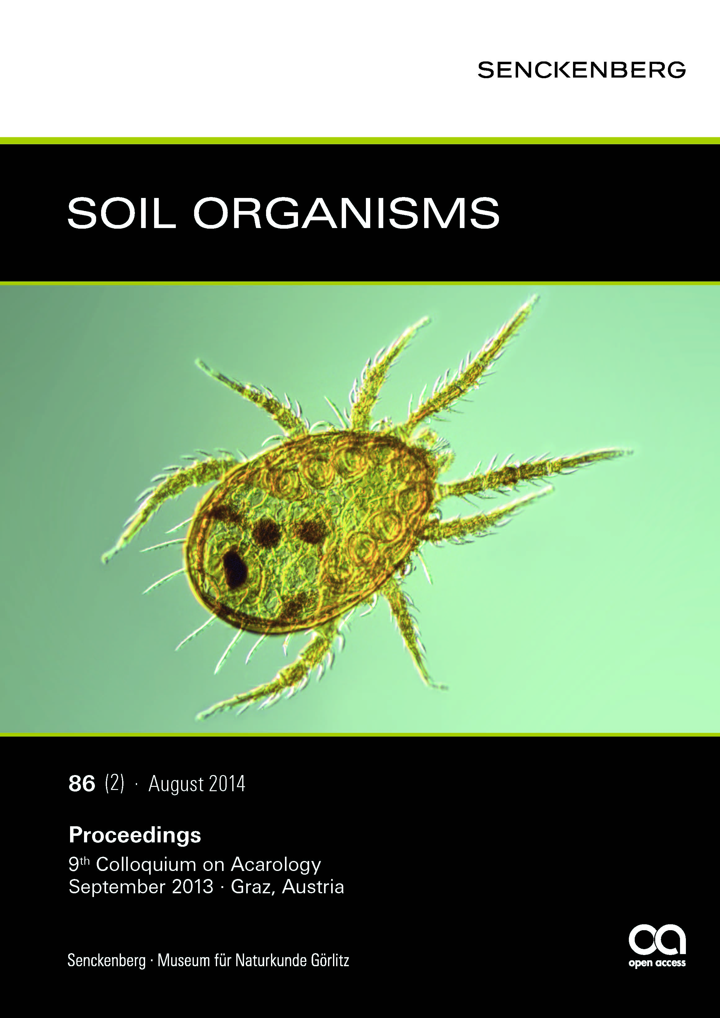
Fine structure of the naso with median eye and trichobothria in the prostigmatid mite Rhagidia halophila(Rhagidiidae, Actinotrichida)
Keywords:
Acari, evolution, ocellus, sensilla, tubular bodies, ultrastructureAbstract
Rhagidia halophila, as other Rhagidiidae, possesses a distinct frontal idiosomatic protuberance, the naso. It bears an unpaired eye (ocellus) that is directed ventrally and consists of four receptor cells provided with numerous rhabdomeric microvilli. The cuticle overlying the microvilli is thin and smooth in contrast to the dorsal cuticle of the naso that shows a fine, spiny sculpture. Details of the fine structure of the receptor cells of the eye are reported. It seems that there is a high membrane turnover which is indicated by numerous dense stacks of membranes. The peculiarity of the median eye and the naso of actinotrichid mites is highlighted and interpreted as plesiomorphic within Arachnida. On the dorsal side of the naso, a pair of small setae (internal verticals) is located in deep sockets thus representing trichobothria. Each sensillum is innervated by two dendrites which terminate with prominent tubular bodies. The axons of the receptor cells of these trichobothria like those of the median eye leave the naso through a narrow passage bordered by specialized cells.
Downloads
Downloads
Published
Issue
Section
License
LicenseSoil Organisms is committed to fair open access publishing. All articles are available online without publication fees. Articles published from Vol. 96 No. 3 (2024) onwards are licensed under the Creative Commons Attribution 4.0 International (CC BY 4.0) license. Articles published from Vol. 80 No. 1 through Vol. 96 No. 2 are available under the previous terms, allowing non-commercial, private, and scientific use.

